Abstract
The brain works based on the complicated electrophysiological activities of the neurons. Many previous researches focused on the measurement of electrophysiological singals of single neurons which were cultured in 2D. However, the neurons always form fascicles in vivo and has a 3D structure in the body. The neural organoid has recently attracted attention from many researchers. The organoid is a simplified version of an organ produced in vitro in 3D, which has similar anatomical characteristics to the tissue in vivo. Although many researches on the techniques to culture the organoids in vitro have been conducted so far, there is no useful mehtod to measure electrophysiologically the whole surface or the inside of a organoid.
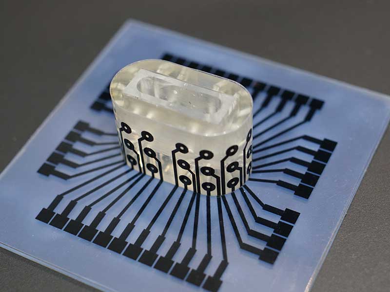
In this study, we are developing the microfluidic device which enables us to culture neurons as a spheroid in 3D and to perform 3D measurement of their electrophysiological activities. The 3D-shaped PDMS device is casted using a plastic mold made by 3D printing. We can culture a spheroid of neurons in the 3D-shaped microfluidic device and measure electrophysiological signals from neurons using the electrodes wired in 3D.
 3D device to measure electrophysiological activities of neurons
Collaboration
- Levi Lab, IIS, University of Tokyo
- Ikeuchi Lab, IIS, University of Tokyo