Abstract
The kidneys in our body filter out the waste solute from blood flow and discharge it from our body as urine. In this kidney function, the ion channels of renal tubular cells have a crucial role. The renal tubular cells have primary cilia which can sense the shear stress caused by urinary flow in a kidney tubule. The primary cilia work to regulate the ion channels and also inhibit cell proliferation in response to the detected shear stress. Although several in vitro studies on the function of the primary cilia have been performed in the past, they have not succeeded in developing primary cilia in vitro except in the serum-starved culture condition and have had a difficulty in observing and evaluating drug responses when the mechanical shear stress is applied to the primary cilia.
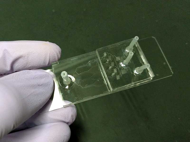 Microfluidic cell culture device for renal tubular cells
Our research group is developing a new microfluidic cell culture system which enables in vitro drug testing of renal tubular cells under the hydrodynamic shear stress. So far, we have successfully cultured renal tubular cells and developed primary cilia in a microfluidic device without requiring any special environmental condition such as the serum starvation. And also the developed microfluidic cell culture system works well to measure the intracellular calcium level which increases in response to the hydrodynamic shear stress level.
Collaboration
- Division of Nephrology and Endocrinology, University of Tokyo Hospital
Sponsor
- AMED